Target of Interest
Search Thermo Fisher Scientific

Clicking the images or links will redirect you to a website hosted by BenchSci that provides third-party scientific content. Neither the content nor the BenchSci technology and processes for selection have been evaluated by us; we are providing them as-is and without warranty of any kind, including for use or application of the Thermo Fisher Scientific products presented.
图: 1 / 3
 Antibody (46-9865-42) in Flow")
 Antibody (46-9865-42) in Flow")
 Antibody (46-9865-42) in Flow")
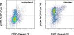
Description: The CR55T33 monoclonal antibody recognizes phosphorylated serine 139 of human and mouse H2AX. H2AX is a member of the H2A histone family that complex with DNA and other histones to form the repeating nucleosome units characteristic of eukaryotic chromatin. Nucleosomes consist of approximately 147 base pairs of DNA wrapped around an octamer of histones composed of two each of the four histone proteins: H2A, H2B, H3 and H4. After induction of DNA damage such as double-strand breaks by irradiation, genotoxic stresses, replication errors or gene recombination, PI3K-like kinases (e.g., ataxia telangiectasia mutated (ATM), ataxia telangiectasia Rad-3-related (ATR), and DNA-dependent protein kinase (DNA-PK) are activated to phosphorylate serine 139 in H2AX. This early phosphorylation event plays a critical role in recruiting proteins involved in DNA repair.
The monoclonal antibody CR55T33 recognizes a single band of approximately 15 kDa on reduced cell lysates from Jurkat cells stimulated with etoposide.
Applications Reported: This CR55T33 antibody has been reported for use in intracellular staining followed by flow cytometric analysis.
Applications Tested: This CR55T33 antibody has been pre-titrated and tested by intracellular staining followed by flow cytometric analysis of treated human peripheral blood cells using the Foxp3/Transcription Factor Buffer Set (Product # 00-5523-00) and protocol . Refer to BestProtocols®; Staining intracellular Antigens for Flow Cytometry Protocol (Protocol B: One step protocol for (nuclear) intracellular proteins). This can be used at 5 µL (0.125 µg) per test. A test is defined as the amount (µg) of antibody that will stain a cell sample in a final volume of 100 µL. Cell number should be determined empirically but can range from 10^5 to 10^8 cells/test.
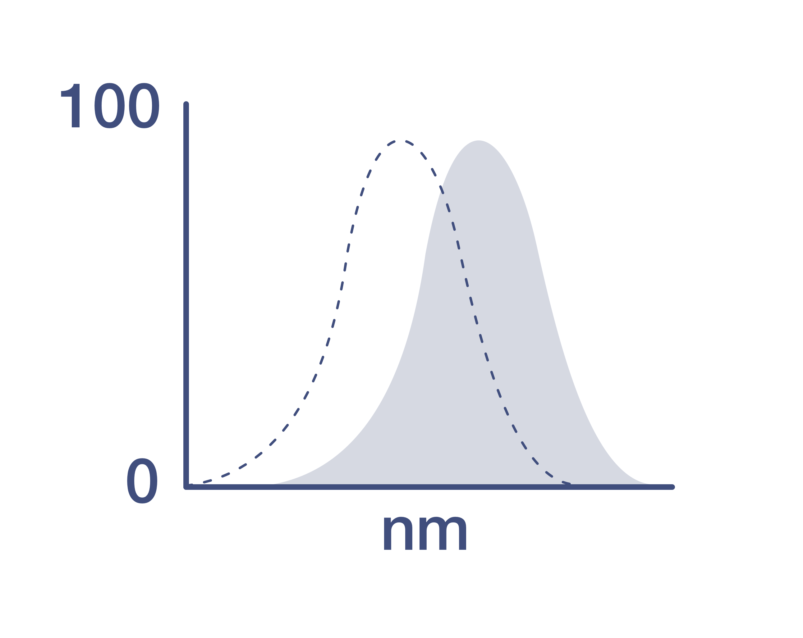
PerCP-eFluor® 710 emits at 710 nm and is excited with the blue laser (488 nm); it can be used in place of PerCP-Cyanine5.5. We recommend using a 710/50 bandpass filter, however, the 695/40 bandpass filter is an acceptable alternative. Please make sure that your instrument is capable of detecting this fluorochrome.
Fixation: Samples can be stored in IC Fixation Buffer (Product # 00-8222) (100 µL of cell sample + 100 µL of IC Fixation Buffer) or 1-step Fix/Lyse Solution (Product # 00-5333) for up to 3 days in the dark at 4°C with minimal impact on brightness and FRET efficiency/compensation. Some generalizations regarding fluorophore performance after fixation can be made, but clone specific performance should be determined empirically.
Excitation: 488 nm; Emission: 710 nm; Laser: Blue Laser.
Filtration: 0.2 µm post-manufacturing filtered.
Histone H2A.X (H2AX) is a member of the histone H2A family which is one of the four core histones making up the nucleosome core particle. In eukaryotes, DNA double strand breaks (DSBs) have been shown to trigger the phosphorylation of serine 139 at the carboxy terminus of histone H2AX resulting in gamma-H2AX. The phosphorylation of H2AX can be detected by Western blotting or immunofluorescence, revealing the frequency of DSBs. The phosphatidylinositol 3-kinases have been implicated in H2AX phosphorylation, but it is unclear if ATM is the primary H2AX kinase or if other members of the family such as DNA-PK and ATR contribute in a similar manner. Structurally, H2A.x contains 143 amino acid residues. Histone H2A.X is considered a basal histone, being synthesized in G1 as well as in S-phase, and its mRNA contains polyA addition motifs and a polyA tail along with the conserved stem-loop and U7 binding sequences involved in the processing and stability of replication type histone mRNAs. There are two forms of Histone H2A.X mRNA, one about 1600 bases long and contains polyA; the other about 575 bases long, lacking polyA. The short form behaves as a replication type histone mRNA, while the longer behaves as a basal type histone mRNA. Histone H2A.X maps to the 11q23.2-q23.3 region of the human chromosome. Histone H2A.x contributes to histone-formation and therefore the structure of DNA. Histone H2A variant H2A.x specifically regulates the interaction of MDC1 (mediator of DNA damage checkpoint protein 1), a DNA repair protein to the sites of DNA damage.
仅用于科研。不用于诊断过程。未经明确授权不得转售。
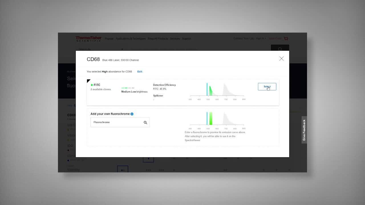
Watch the video to learn how to use the Invitrogen Flow Cytometry Panel Builder to build your next flow cytometry panel in 5 easy steps.
蛋白别名: gamma H2AX; H2A histone family, member X; H2a/x; H2AX histone; histone 5 protein 2ax; Histone H2A.X; Histone H2a/x; Histone H2AX
基因别名: AW228881; gammaH2ax; H2A.X; H2A/X; H2AFX; H2AX; Hist5-2ax
UniProt ID: (Human) P16104, (Mouse) P27661
Entrez Gene ID: (Human) 3014, (Mouse) 15270

