 Shujia Zhu, principal investigator at the Institute of Neuroscience, Chinese Academy of Sciences, says cryo-electron microscopy (cryo-EM) is fundamental for collaboration across research disciplines and drug discovery.
When asked about the future of this technique, especially in neuroscience research, she said “tomography is the future.”
Can you tell us about your work in neuroscience research?
I have been fascinated by the physiological role of the NMDA receptors for almost 15 years. When I was a master’s student, I mainly used animal models with genetic modified NMDA receptors to study learning and memory. When I finished my master’s, I found some of those behavior experiments difficult to reproduce. So, I decided to pursue studies of the NMDA receptors on the molecular level and know more about the biophysical property of these receptors.
I moved to France and pursued my PhD using electrophysiological and functional tools to study gating mechanisms of the NMDA receptors. When I finished my PhD, I found functional data difficult to interpret without the structural insights. Thus, I moved to the states for my postdoc training and tried to elucidate the structure of the NMDA receptors. During this time, from 2014 to 2016, there was a breakthrough in cryo-EM called the resolution revolution. Before my post doc, I didn’t even know how to do protein purification. I hadn’t done any structural work yet. With the help of cryo-EM, I finished my post doc training within two years and got a bunch of new NMDA structures in biological-relevant states. Then, I got a group leader position at the Institute of Neuroscience in China.
You see, I jumped from animal model to molecular layers to structure. In my lab, I try to combine all these research aspects together. We can do structure, we can do function, and we can also study animal behavior.
I would like to understand the function of various NMDA receptors from the atomic structural resolution, from the molecular gating mechanism, as well as from the brain circuit level.
What success have you experienced using cryo-EM for neuroscience research?
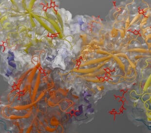
I can share two success stories with you. The first is about our paper, Gating mechanism and a modulatory niche of human GluN1-GluN2A NMDA receptors.
For this project, we wanted to understand the molecular insights on the gating mechanism in GluN1-GluN2A subtype NMDA receptors. The channel activity of NMDA receptor could be precisely modulated by endogenous or exogenous ligands. We wanted to understand how the channel was inhibited, activated, or regulated by allosteric modulator and channel blockers. In combining cryo-EM with pharmacological tools, we got a gallery of NMDA receptor structures captured in the inhibited, the activated, and the modulated states. The most interesting part is that we discovered a new binding site for the molecule called the 9-Aminoacridine. The cryo-EM density showed that 9-Aminoacridine resides at the LBD-TMD linker cavity, which has never been reported as a ligand binding pocket in the field of NMDA receptors. This study enriched the understandings of NMDA receptor structure and function, unraveled gating mechanism and provided molecular evidence for drug design and development.
It’s almost impossible to achieve these accomplishments by x-ray crystallography.

Shujia Zhu conducting neuroscience research with students at Chinese Academy of Sciences
Another story was published in July 2021 (Structural basis of ketamine action on human NMDA receptors).
In this study, we focused on a rapid antidepressant drug called S-ketamine. Depression (major depressive disorder) affects more than 10% of world population. Traditional antidepressants typically take weeks or even months to take affect and 1/3 of patients are (treatment resistant) to traditional methods. In the field, people know that S-ketamine is a pore blocker of the NMDA receptors, but nobody has visualized where ketamine binds and how it inhibits NMDA receptor channels. We determined the cryo-EM structures of human GluN1-GluN2A and GluN1-GluN2B NMDA receptors in complex with S-ketamine and found the electron density of S-ketamine in the central vestibule of transmembrane domain in both receptors. Combining with in silico calculation and electrophysiology, we revealed that two amino acids, L642 in GluN2A and N616 in GluN1, were the key residues forming hydrophobic interaction and hydrogen bond with S-ketamine. I think our discovery will pave the way for the future development of rapid antidepressant targeting to the glutamate receptors.
What will the next few years look like for you and your lab?
We are mainly doing single particle analysis in our lab right now, but I think tomography is the future.
We do single particle analysis using receptors expressed by recombinant expression systems, but we really would like to understand how the receptors look and function in native brain tissue. I think we will move to tomography in the next five years and I’m currently learning that.
How prevalent is cryo-EM in China? Why is it important for research institutions to invest in cryo-EM?
It is the best time to do research in China. We have great students, great post docs, and great collaboration. Both the government and private sectors are investing in cryo-EM.
Cryo-EM is accelerating basic research and drug discovery. I think cryo-EM is the first step in integrating different disciplines like biochemistry, electrophysiology, computational analysis. We use this fundamental technique to get the structure then we can initiate or accelerate other collaborations.
//
Suzanne Graham is the Senior Director Business Development at Thermo Fisher Scientific.
Subscribe Now to receive new Accelerating Microscopy posts straight to your inbox.
Guiding the Development of an HIV Vaccine with Structure-Based Drug Design
Novel drug development in the fight against the HIV pandemic...
Read More
Virus microscopy unravels chikungunya virus structure
Global impacts of the chikungunya virus Chikungunya is a vir...
Read More
What is cryo-EM?
Cryo-EM: cryo electron microscopy Cryo-electron microscopy (...
Read More
Glacios 2 Cryo-TEM and the Evolution of 200 kV Cryo-EM
Glacios 2 Cryo-TEM and the Evolution of 200 kV Cryo-EM Cryo-...
Read More
Leave a Reply