Search
Disclaimer

Clicking the images or links will redirect you to a website hosted by BenchSci that provides third-party scientific content. Neither the content nor the BenchSci technology and processes for selection have been evaluated by us; we are providing them as-is and without warranty of any kind, including for use or application of the Thermo Fisher Scientific products presented.
Invitrogen
BrdU Monoclonal Antibody (BU20A), PerCP-eFluor™ 710, eBioscience™
{{$productOrderCtrl.translations['antibody.pdp.commerceCard.promotion.promotions']}}
{{$productOrderCtrl.translations['antibody.pdp.commerceCard.promotion.viewpromo']}}
{{$productOrderCtrl.translations['antibody.pdp.commerceCard.promotion.promocode']}}: {{promo.promoCode}} {{promo.promoTitle}} {{promo.promoDescription}}. {{$productOrderCtrl.translations['antibody.pdp.commerceCard.promotion.learnmore']}}
")
图: 1 / 15
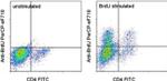
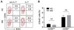
BrdU Antibody (46-5071-42) in Flow
Anti-CD3/CD28 (Product # 16-0031-82, 16-0281)-stimulated mouse splenocytes either unlabeled (left) or labeled with BrdU (right) were surface stained with Anti-Mouse CD4 FITC (Product # 11-0041-82). These cells were then stained intracellularly with Anti-BrdU PerCP-eFluor® 710 using the BrdU Staining Kit for Flow Cytometry PerCP-eFluor® 710 and protocol. Total viable cells were used for analysis.

 in Flow")
 in Flow")
 in Flow")
 in Flow")
 in Flow")
 in Flow")
 in Flow")
 in Flow")
 in Flow")
 in Flow")
 in Flow")
 in Flow")
 in Flow")
 in Flow")
 in Flow")
产品信息
46-5071-42
应用
建议稀释比
已发表文章
产品规格
种属反应
Chemical
已发表种属
Chemical
宿主/亚型
Mouse
/ IgG1, kappa
分类
Monoclonal
类型
Antibody
克隆号
BU20A
偶联物
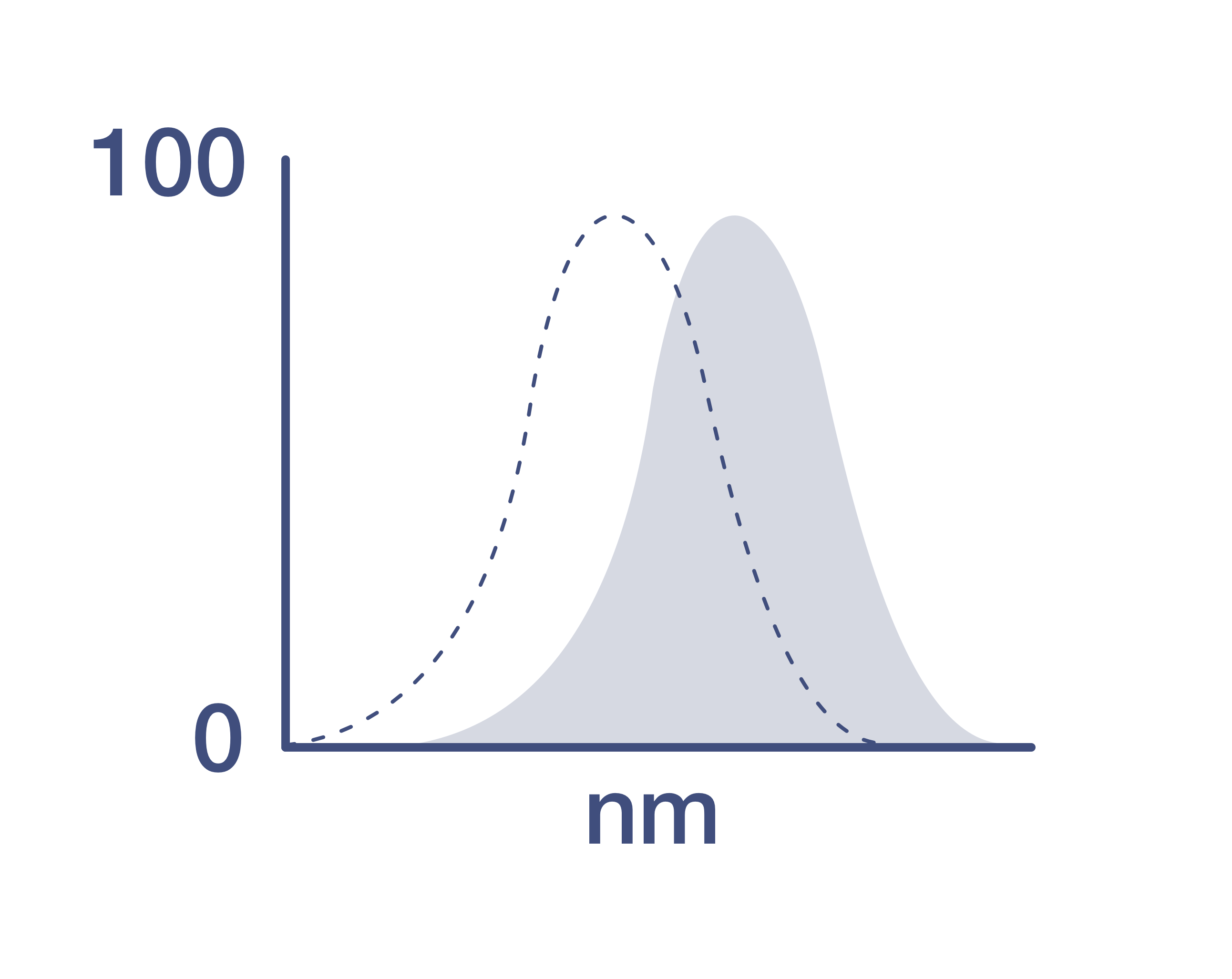
激发/发射光谱
482/708 nm
查看光谱
形式
Liquid
浓度
5 µL/Test
纯化类型
Affinity chromatography
保存液
PBS, pH 7.2, with BSA
内含物
0.09% sodium azide
保存条件
4°C, store in dark, DO NOT FREEZE!
运输条件
Ambient (domestic); Wet ice (international)
RRID
产品详细信息
Description: This Bu20a monoclonal antibody reacts with 5-bromodeoxyuridine (BrdU). BrdU is a derivative of uridine that can be incorporated into DNA in place of thymidine during the S-phase of the cell cycle. Anti-BrdU can then be used to identify cells that have undergone DNA synthesis during BrdU treatment.
For staining for flow cytometric analysis, we recommend the use of the BrdU Staining Buffer Set (Product # 00-5525) and protocol.
Applications Reported: This BU20A antibody has been reported for use in intracellular staining followed by flow cytometric analysis.
Applications Tested: This BU20A antibody has been pre-titrated and tested by flow cytometric analysis of BrdU-pulsed mouse splenocytes using the Foxp3/Transcription Factor Buffer Set (Product # 00-5523-00) and protocol or the BrdU Staining Buffer Set (Product # 00-5525-00) and protocol. This can be used at 5 µL (0.06 µg) per test. A test is defined as the amount (µg) of antibody that will stain a cell sample in a final volume of 100 µL. Cell number should be determined empirically but can range from 10^5 to 10^8 cells/test.
PerCP-eFluor® 710 can be used in place of PE-Cy5, PE-Cy5.5 or PerCP-Cy5.5. PerCP-eFluor® 710 emits at 710 nm and is excited with the blue laser (488 nm). Please make sure that your instrument is capable of detecting this fluorochrome. For a filter configuration, we recommend using the 685 LP dichroic mirror and 710/40 band pass filter, however the 695/40 band pass filter is an acceptable alternative.
Our testing indicates that PerCP-eFluor® 710 conjugated antibodies are stable when stained samples are exposed to freshly prepared 2% formaldehyde overnight at 4°C, but please evaluate for alternative fixation protocols. BrdU labeling and staining with the Anti-BrdU antibody:1. Label dividing cells with 10 µM BrdU for 45 min at 37°C.2. Following the incubation, harvest the cells and wash once with 1X PBS.3. Stain surface molecules according to the Surface Staining Protocol.4. Wash in cold Flow Cytometry Staining Buffer or 1X PBS.5. Resuspend the cell pellet by pulse vortexing. Then add 1 mL of freshly prepared Foxp3 Fixation/Permeabilization Buffer (Product # 00-5521) to each sample. pulse vortex again.6. Incubate for 30 to 60 minutes at 2-8°C in the dark.7. Wash once with cold Flow Cytometry Staining Buffer followed by centrifugation. Decant the supernatant.8. Resuspend the cell pellet with 100 µL Flow Cytometry Staining Buffer containing 30 µg of Dnase I.9. Incubate for 1 hr at 37°C and then wash.10. Stain cells with anti-BrdU antibody for 30 min to 1 hr and then wash.10. Analyze the samples.
Excitation: 488 nm; Emission: 710 nm; Laser: Blue Laser.
Filtration: 0.2 µm post-manufacturing filtered.
靶标信息
Bromodeoxyuridine (5-bromo-2-deoxyuridine, BrdU) is a synthetic nucleoside that is an analogue of thymidine. BrdU is commonly used in the detection of proliferating cells in living tissues, and can be incorporated into the newly synthesized DNA of replicating cells (during the S phase of the cell cycle), substituting for thymidine during DNA replication. Antibodies specific for BrdU can then be used to detect the incorporated chemical, thus indicating cells that were actively replicating their DNA. Binding of the antibody requires denaturation of the DNA, usually by exposing the cells to acid or heat.
仅用于科研。不用于诊断过程。未经明确授权不得转售。
How to use the Panel Builder
Watch the video to learn how to use the Invitrogen Flow Cytometry Panel Builder to build your next flow cytometry panel in 5 easy steps.

