Search
Disclaimer

Clicking the images or links will redirect you to a website hosted by BenchSci that provides third-party scientific content. Neither the content nor the BenchSci technology and processes for selection have been evaluated by us; we are providing them as-is and without warranty of any kind, including for use or application of the Thermo Fisher Scientific products presented.
Invitrogen
CD8a Monoclonal Antibody (SK1), NovaFluor™ Yellow 755, eBioscience™
{{$productOrderCtrl.translations['antibody.pdp.commerceCard.promotion.promotions']}}
{{$productOrderCtrl.translations['antibody.pdp.commerceCard.promotion.viewpromo']}}
{{$productOrderCtrl.translations['antibody.pdp.commerceCard.promotion.promocode']}}: {{promo.promoCode}} {{promo.promoTitle}} {{promo.promoDescription}}. {{$productOrderCtrl.translations['antibody.pdp.commerceCard.promotion.learnmore']}}
")
图: 1 / 2
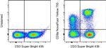
CD8a Antibody (H023T03Y08) in Flow
Normal human peripheral blood cells were unstained (left) or stained with CD8a Monoclonal Antibody, NovaFluor Yellow 755 (Product # H023T03Y08) (right). All cells were co-stained with CD3 Monoclonal Antibody, eFluor 450 (Product # 62-0038-42). Total viable cells in the lymphocyte gate were used for analysis, as determined by LIVE/DEAD Blue (Product # L34962). Data was acquired on a 5-laser Cytek Aurora and unmixed with autofluorescence extraction.
 in Flow")
 in Flow")
产品信息
H023T03Y08
产品规格
宿主/亚型
Mouse
/ IgG1, kappa
分类
Monoclonal
类型
Antibody
克隆号
SK1
偶联物
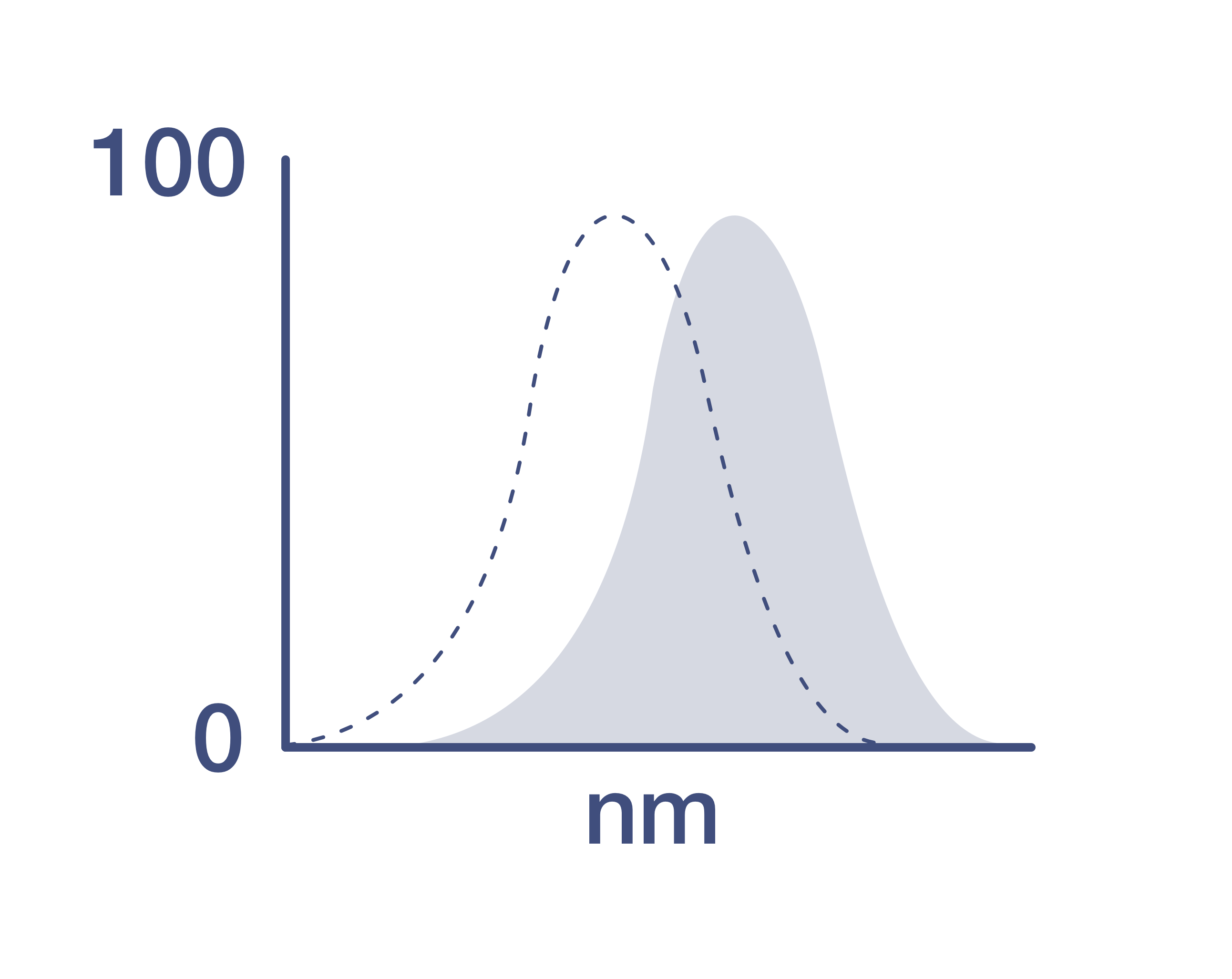
激发/发射光谱
551/755 nm
查看光谱
形式
Liquid
浓度
0.6 µg/Test
保存条件
4°C, store in dark, DO NOT FREEZE!
运输条件
Ambient (domestic); Wet ice (international)
产品详细信息
Description: The SK1 monoclonal antibody reacts with the human CD8a molecule, an approximately 32-34 kDa cell surface receptor expressed either as a heterodimer with the CD8 beta chain (CD8 alpha/beta) or as a homodimer (CD8 alpha/alpha). A majority of thymocytes and a subpopulation of mature T cells and NK cells express CD8a. CD8 binds to MHC class I and through its association with protein tyrosine kinase p56lck plays a role in T-cell development and activation of mature T cells.
Applications Reported: This SK1 antibody has been reported for use in flow cytometric analysis.
Applications Tested: This SK1 antibody has been pre-titrated and tested by flow cytometric analysis of normal human peripheral blood cells. This can be used at 5 µL (0.06 µg) per test. A test is defined as the amount (µg) of antibody that will stain a cell sample in a final volume of 100 µL. Cell number should be determined empirically but can range from 10^5 to 10^8 cells/test.
NovaFluor dyes are not compatible with DNA intercalating viability dyes. Do not use viability dyes such as propidium iodide, 7-actinomycin D (7-AAD) and DAPI. Invitrogen LIVE/DEAD Fixable Dead Cell stains are recommended for use with NovaFluor dyes.
Each NovaFluor conjugate or kit is shipped with CellBlox Blocking Buffer. Use this buffer whenever staining with NovaFluor conjugates, including single-color compensation controls using cells. Whenever possible, we recommend adding CellBlox Blocking Buffer to antibody cocktails/master mixes prior to combining with cells. Add 5 µL per sample (regardless of the number of NovaFluors in your panel) to use the antibody cocktail as intended. For single-color controls, use 5 µL of CellBlox Blocking Buffer per 100µL of cell sample containing 10^3 to 10^8 cells.
NovaFluor conjugates are based on Phiton™ technology utilizing novel nucleic acid dye structures that allow for engineered fluorescent signatures with consideration for spillover and spread impacts. Learn more
Excitation: 552 nm; Emission: 755 nm; Laser: 561 nm (Yellow) Laser
靶标信息
CD8, also known as cluster of differentiation 8, is a type I transmembrane glycoprotein of the immunoglobulin family that plays a crucial role in T cell differentiation, activation, and signal transduction. It is expressed as either a heterodimer (CD8 alpha beta) or a homodimer (CD8 alpha alpha). The CD8 alpha beta form is predominantly found on the majority of thymocytes and a subpopulation of mature alpha beta TCR T cells, while the CD8 alpha alpha form is expressed on gamma delta TCR T cells, a subset of intestinal intraepithelial lymphocytes (IELs), and dendritic cells. CD8 functions as a co-receptor for major histocompatibility complex class I (MHC-I) molecules, working alongside the T cell receptor (TCR). The CD8 alpha chain is essential for binding to MHC-I. CD8 is also expressed on a subset of T cells, NK cells, monocytes, and dendritic cells as disulfide-linked homodimers of CD8 alpha. Upon ligation of MHC-I/peptide complexes presented by antigen-presenting cells (APCs), CD8 recruits lymphocyte-specific protein tyrosine kinase (Lck), leading to lymphokine production, increased motility, and activation of cytotoxic T lymphocytes (CTLs). Activated CTLs are vital for clearing pathogens and tumor cells. The differentiation of naive CD8+ T cells into CTLs is strongly enhanced by cytokines such as IL-2, IL-12, and TGF-beta1. Through its interactions with MHC-I and association with protein tyrosine kinase p56lck, CD8 plays a significant role in T cell development and the activation of mature T cells.
仅用于科研。不用于诊断过程。未经明确授权不得转售。
How to use the Panel Builder
Watch the video to learn how to use the Invitrogen Flow Cytometry Panel Builder to build your next flow cytometry panel in 5 easy steps.
篇参考文献 (0)
您是否在文献中引用过该产品?请点击下方按钮邮件告知我们。

