Search
Disclaimer

Clicking the images or links will redirect you to a website hosted by BenchSci that provides third-party scientific content. Neither the content nor the BenchSci technology and processes for selection have been evaluated by us; we are providing them as-is and without warranty of any kind, including for use or application of the Thermo Fisher Scientific products presented.
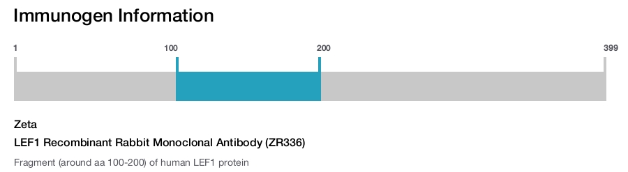
Zeta
LEF1 Recombinant Rabbit Monoclonal Antibody (ZR336)
{{$productOrderCtrl.translations['antibody.pdp.commerceCard.promotion.promotions']}}
{{$productOrderCtrl.translations['antibody.pdp.commerceCard.promotion.viewpromo']}}
{{$productOrderCtrl.translations['antibody.pdp.commerceCard.promotion.promocode']}}: {{promo.promoCode}} {{promo.promoTitle}} {{promo.promoDescription}}. {{$productOrderCtrl.translations['antibody.pdp.commerceCard.promotion.learnmore']}}
 (IHC (P))")
 in IHC (P)")
产品信息
Z2642RP
产品规格
种属反应
Human
宿主/亚型
Rabbit
/ IgG
Expression System
CHO cells
分类
Recombinant Monoclonal
类型
Antibody
克隆号
ZR336
抗原
Fragment (around aa 100-200) of human LEF1 protein
偶联物
形式
Liquid
纯化类型
Protein A
保存液
tris with BSA, NP-40
内含物
<0.1% sodium azide
保存条件
4°C
运输条件
Ambient (domestic); Wet ice (international)
产品详细信息
This product is diluted and in a ready-to-use formulation.
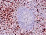
A recommended positive control tissue for this product is Tonsil (B cells), however positive controls are not limited to this tissue type.
The primary antibody is intended for laboratory professional use in the detection of the corresponding protein in formalin-fixed, paraffin-embedded tissue stained in manual qualitative immunohistochemistry (IHC) testing. This antibody is intended to be used after the primary diagnosis of tumor has been made by conventional histopathology using non-immunological histochemical stains.
Lymphoid Enhancing Factor 1 (LEF1) is a transcription factor that belongs to the TCF/LEF family. LEF1 participates as a regulator in Wnt signaling pathways. LEF1 is an important factor in lymphopoiesis and is expressed normally in T and pro-B cells but not expressed in mature B cells. Strong nuclear expression of LEF1 has been observed in majority of chronic lymphocytic leukemia/small lymphocytic lymphoma cases and LEF1 is not detected in other small B cell lymphomas. Anti-LEF1 may be used as an aid for differentiation of chronic lymphocytic leukemia/small lymphocytic lymphoma from other small B cell lymphomas.
Antibody is used with formalin-fixed and paraffin-embedded sections. Pretreatment of deparaffinized tissue with heat-induced epitope retrieval or enzymatic retrieval is recommended. In general, immunohistochemical (IHC) staining techniques allow for the visualization of antigens via the sequential application of a specific antibody to the antigen (primary antibody), a secondary antibody to the primary antibody (link antibody), an enzyme complex and a chromogenic substrate with interposed washing steps. The enzymatic activation of the chromogen results in a visible reaction product at the antigen site. Results are interpreted using a light microscope and aid in the differential diagnosis of pathophysiological processes, which may or may not be associated with a particular antigen.
A positive tissue control must be run with every staining procedure performed. This tissue may contain both positive and negative staining cells or tissue components and serve as both the positive and negative control tissue. External Positive control materials should be fresh autopsy/biopsy/surgical specimens fixed, processed and embedded as soon as possible in the same manner as the patient sample (s). Positive tissue controls are indicative of correctly prepared tissues and proper staining methods. The tissues used for the external positive control materials should be selected from the patient specimens with well-characterized low levels of the positive target activity that gives weak positive staining. The low level of positivity for external positive controls is designed to ensure detection of subtle changes in the primary antibody sensitivity from instability or problems with the staining methodology. A tissue with weak positive staining is more suitable for optimal quality control and for detecting minor levels of reagent degradation.
Internal or external negative control tissue may be used depending on the guidelines and policies that govern the organization to which the end user belongs to. The variety of cell types present in many tissue sections offers internal negative control sites, but this should be verified by the user. The components that do not stain should demonstrate the absence of specific staining, and provide an indication of non-specific background staining. If specific staining occurs in the negative tissue control sites, results with the patient specimens must be considered invalid.
靶标信息
This gene encodes a transcription factor belonging to a family of proteins that share homology with the high mobility group protein-1. The protein encoded by this gene can bind to a functionally important site in the T-cell receptor-alpha enhancer, thereby conferring maximal enhancer activity. This transcription factor is involved in the Wnt signaling pathway, and it may function in hair cell differentiation and follicle morphogenesis. Mutations in this gene have been found in somatic sebaceous tumors. This gene has also been linked to other cancers, including androgen-independent prostate cancer. Alternative splicing results in multiple transcript variants.
仅用于科研。不用于诊断过程。未经明确授权不得转售。
篇参考文献 (0)
您是否在文献中引用过该产品?请点击下方按钮邮件告知我们。
生物信息学
蛋白别名: DKFZp586H0919; FLJ46390; HGNC:6551; HMG Binding Domain; LEF-1; Lymphoid enhancer-binding factor 1; OTTHUMP00000219736; OTTHUMP00000219737; T cell-specific transcription factor 1-alpha; TCF1-alpha
基因别名: LEF-1; LEF1; TCF10; TCF1ALPHA; TCF7L3
UniProt ID: (Human) Q9UJU2
Entrez Gene ID: (Human) 51176
RNA polymerase II regulatory region sequence-specific DNA binding
RNA polymerase II core promoter proximal region sequence-specific DNA binding
transcriptional activator activity, RNA polymerase II core promoter proximal region sequence-specific binding
transcriptional activator activity, RNA polymerase II transcription regulatory region sequence-specific binding
DNA binding
chromatin binding
transcription factor activity, sequence-specific DNA binding
transcription factor activity, RNA polymerase II distal enhancer sequence-specific binding
protein binding
beta-catenin binding
transcription factor binding
DNA binding, bending
estrogen receptor activity
estrogen receptor binding
enhancer binding
histone binding
histone deacetylase binding
cysteine-type endopeptidase inhibitor activity involved in apoptotic process
sequence-specific DNA binding
transcription regulatory region DNA binding
gamma-catenin binding
armadillo repeat domain binding
C2H2 zinc finger domain binding
DNA-binding transcription factor
gene-specific transcriptional regulator
negative regulation of transcription from RNA polymerase II promoter
patterning of blood vessels
osteoblast differentiation
neural crest cell migration
somitogenesis
kidney development
epithelial to mesenchymal transition
sprouting angiogenesis
transcription from RNA polymerase II promoter
Wnt signaling pathway, calcium modulating pathway
positive regulation of cell proliferation
response to lithium ion
positive regulation of gene expression
positive regulation of epithelial to mesenchymal transition
Wnt signaling pathway
regulation of striated muscle tissue development
dentate gyrus development
hypothalamus development
forebrain radial glial cell differentiation
forebrain neuroblast division
forebrain neuron differentiation
formation of radial glial scaffolds
regulation of cell-cell adhesion
neutrophil differentiation
positive regulation of cell growth
embryonic limb morphogenesis
positive regulation of cell migration
BMP signaling pathway
positive regulation of granulocyte differentiation
mammary gland development
organ regeneration
negative regulation of interleukin-13 production
negative regulation of interleukin-4 production
negative regulation of interleukin-5 production
T cell receptor V(D)J recombination
B cell proliferation
odontogenesis of dentin-containing tooth
negative regulation of apoptotic process
negative regulation of cysteine-type endopeptidase activity involved in apoptotic process
negative regulation of DNA binding
steroid hormone mediated signaling pathway
tongue development
skin development
positive regulation by host of viral transcription
histone H3 acetylation
histone H4 acetylation
T-helper 1 cell differentiation
negative regulation of striated muscle tissue development
negative regulation of transcription, DNA-templated
positive regulation of transcription, DNA-templated
positive regulation of transcription from RNA polymerase II promoter
alpha-beta T cell differentiation
eye pigmentation
paraxial mesoderm formation
muscle fiber development
sensory perception of taste
palate development
anatomical structure regression
canonical Wnt signaling pathway
face morphogenesis
cell chemotaxis
apoptotic process involved in morphogenesis
chorio-allantoic fusion
trachea gland development
cellular response to cytokine stimulus
cellular response to interleukin-4
positive regulation of cell proliferation in bone marrow
negative regulation of apoptotic process in bone marrow
odontoblast differentiation
negative regulation of estrogen receptor binding
negative regulation of canonical Wnt signaling pathway
apoptotic process involved in patterning of blood vessels
beta-catenin-TCF complex assembly

