Target of Interest
Search Thermo Fisher Scientific

Clicking the images or links will redirect you to a website hosted by BenchSci that provides third-party scientific content. Neither the content nor the BenchSci technology and processes for selection have been evaluated by us; we are providing them as-is and without warranty of any kind, including for use or application of the Thermo Fisher Scientific products presented.
 in IHC (P)")
This product is diluted and in a ready-to-use formulation.
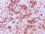
A recommended positive control tissue for this product is Melanoma, however positive controls are not limited to this tissue type.
The primary antibody is intended for laboratory professional use in the detection of the corresponding protein in formalin-fixed, paraffin-embedded tissue stained in manual qualitative immunohistochemistry (IHC) testing. This antibody is intended to be used after the primary diagnosis of tumor has been made by conventional histopathology using non-immunological histochemical stains.
Tyrosinase is a copper-containing metalloglycoprotein that catalyzes several steps in the melanin pigment biosynthetic pathway; the hydroxylation of tyrosine to L-3,4-dihydroxyphenylalanine (dopa), and the subsequent oxidation of dopa to dopaquinone. Mutations of the tyrosinase gene occur in various forms of albinism. Tyrosinase is one of the targets for cytotoxic T-cell recognition in melanoma patients. Clone T311 shows no cross-reaction with MAGE-1 and tyrosinase-related protein 1, TRP-1/gp75.1 Staining of melanomas with clone T311 showed tyrosinase in melanotic as well as amelanotic variants.1 Ab-1 is a useful marker for melanocytes and melanomas. Occasionally a minor band at 55 kDa is also detected in Western Blotting.
Antibody is used with formalin-fixed and paraffin-embedded sections. Pretreatment of deparaffinized tissue with heat-induced epitope retrieval or enzymatic retrieval is recommended. In general, immunohistochemical (IHC) staining techniques allow for the visualization of antigens via the sequential application of a specific antibody to the antigen (primary antibody), a secondary antibody to the primary antibody (link antibody), an enzyme complex and a chromogenic substrate with interposed washing steps. The enzymatic activation of the chromogen results in a visible reaction product at the antigen site. Results are interpreted using a light microscope and aid in the differential diagnosis of pathophysiological processes, which may or may not be associated with a particular antigen.
A positive tissue control must be run with every staining procedure performed. This tissue may contain both positive and negative staining cells or tissue components and serve as both the positive and negative control tissue. External Positive control materials should be fresh autopsy/biopsy/surgical specimens fixed, processed and embedded as soon as possible in the same manner as the patient sample (s). Positive tissue controls are indicative of correctly prepared tissues and proper staining methods. The tissues used for the external positive control materials should be selected from the patient specimens with well-characterized low levels of the positive target activity that gives weak positive staining. The low level of positivity for external positive controls is designed to ensure detection of subtle changes in the primary antibody sensitivity from instability or problems with the staining methodology. A tissue with weak positive staining is more suitable for optimal quality control and for detecting minor levels of reagent degradation.
Internal or external negative control tissue may be used depending on the guidelines and policies that govern the organization to which the end user belongs to. The variety of cell types present in many tissue sections offers internal negative control sites, but this should be verified by the user. The components that do not stain should demonstrate the absence of specific staining, and provide an indication of non-specific background staining. If specific staining occurs in the negative tissue control sites, results with the patient specimens must be considered invalid.
The enzyme encoded by this gene catalyzes the first 2 steps, and at least 1 subsequent step, in the conversion of tyrosine to melanin. The enzyme has both tyrosine hydroxylase and dopa oxidase catalytic activities, and requires copper for function. Mutations in this gene result in oculocutaneous albinism, and nonpathologic polymorphisms result in skin pigmentation variation. The human genome contains a pseudogene similar to the 3' half of this gene.
仅用于科研。不用于诊断过程。未经明确授权不得转售。
蛋白别名: LB24-AB; Monophenol monooxygenase; oculocutaneous albinism IA; SK29-AB; Tumor rejection antigen AB; Tyrosinase
基因别名: ATN; CMM8; OCA1; OCA1A; OCAIA; SHEP3; TYR
UniProt ID: (Human) P14679
Entrez Gene ID: (Human) 7299