Search
Disclaimer

Clicking the images or links will redirect you to a website hosted by BenchSci that provides third-party scientific content. Neither the content nor the BenchSci technology and processes for selection have been evaluated by us; we are providing them as-is and without warranty of any kind, including for use or application of the Thermo Fisher Scientific products presented.
Zeta
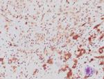
p120 Recombinant Rabbit Monoclonal Antibody (ZR316), RAbMono™
{{$productOrderCtrl.translations['antibody.pdp.commerceCard.promotion.promotions']}}
{{$productOrderCtrl.translations['antibody.pdp.commerceCard.promotion.viewpromo']}}
{{$productOrderCtrl.translations['antibody.pdp.commerceCard.promotion.promocode']}}: {{promo.promoCode}} {{promo.promoTitle}} {{promo.promoDescription}}. {{$productOrderCtrl.translations['antibody.pdp.commerceCard.promotion.learnmore']}}
 (IHC (P))")
 in IHC (P)")
产品信息
Z2620RS
产品规格
种属反应
Human
宿主/亚型
Rabbit
/ IgG
Expression System
CHO cells
分类
Recombinant Monoclonal
类型
Antibody
克隆号
ZR316
抗原
Fragment (around aa 900-1,000) of human CTNND1 protein
偶联物
形式
Liquid
浓度
200 µg/mL
纯化类型
Protein A
保存液
tris with BSA, NP-40
内含物
<0.1% sodium azide
保存条件
4°C
运输条件
Ambient (domestic); Wet ice (international)
产品详细信息
A recommended positive control tissue for this product is breast lobular carcinoma, however positive controls are not limited to this tissue type.
The primary antibody is intended for laboratory professional use in the detection of the corresponding protein in formalin-fixed, paraffin-embedded tissue stained in manual qualitative immunohistochemistry (IHC) testing. This antibody is intended to be used after the primary diagnosis of tumor has been made by conventional histopathology using non-immunological histochemical stains.
Alpha-catenin and beta-catenin bind to the intracellular domain of E-cadherin while p120 catenin binds E-cadherin at a juxta-membrane site. The complex stabilizes tight junctions. In the cell, p120 catenin localized to the E-cadherin/catenins cell adhesion complex, directly associates with cytoplasmic C-terminus of E-cadherin and may similarly interact with other cadherins. p120 is a proliferation-associated nucleolar protein found in most human malignant tumors, but not in resting normal cells. In colorectal cancer the altered localization of p120 catenin corresponds with loss of cytoplasmic localization of E-cadherin. Studies have shown accurate categorization of ductal vs. lobular neoplasia in the breast was achieved with p120 staining. p120 expression further clarifies the separation of low-grade ductal carcinoma in situ from lobular neoplasia. Studies also have shown that altered expression of p120 catenin antibody predicts poor outcome in invasive breast cancer.
Antibody is used with formalin-fixed and paraffin-embedded sections. Pretreatment of deparaffinized tissue with heat-induced epitope retrieval or enzymatic retrieval is recommended. In general, immunohistochemical (IHC) staining techniques allow for the visualization of antigens via the sequential application of a specific antibody to the antigen (primary antibody), a secondary antibody to the primary antibody (link antibody), an enzyme complex and a chromogenic substrate with interposed washing steps. The enzymatic activation of the chromogen results in a visible reaction product at the antigen site. Results are interpreted using a light microscope and aid in the differential diagnosis of pathophysiological processes, which may or may not be associated with a particular antigen.
A positive tissue control must be run with every staining procedure performed. This tissue may contain both positive and negative staining cells or tissue components and serve as both the positive and negative control tissue. External Positive control materials should be fresh autopsy/biopsy/surgical specimens fixed, processed and embedded as soon as possible in the same manner as the patient sample (s). Positive tissue controls are indicative of correctly prepared tissues and proper staining methods. The tissues used for the external positive control materials should be selected from the patient specimens with well-characterized low levels of the positive target activity that gives weak positive staining. The low level of positivity for external positive controls is designed to ensure detection of subtle changes in the primary antibody sensitivity from instability or problems with the staining methodology. A tissue with weak positive staining is more suitable for optimal quality control and for detecting minor levels of reagent degradation.
Internal or external negative control tissue may be used depending on the guidelines and policies that govern the organization to which the end user belongs to. The variety of cell types present in many tissue sections offers internal negative control sites, but this should be verified by the user. The components that do not stain should demonstrate the absence of specific staining, and provide an indication of non-specific background staining. If specific staining occurs in the negative tissue control sites, results with the patient specimens must be considered invalid.
靶标信息
Delta 1 Catenin is an efficient tyrosine kinase substrate implicated both in cell transformation by SRC and in ligand-induced receptor signaling through the EGF, PDGF, CSF-1 and ERBB2 receptors. The association of catenins to cadherins produces a complex which is linked to the actin filament network, and which seems to be of primary importance for cadherins cell-adhesion properties.
仅用于科研。不用于诊断过程。未经明确授权不得转售。
篇参考文献 (0)
您是否在文献中引用过该产品?请点击下方按钮邮件告知我们。
生物信息学
蛋白别名: Cadherin-associated Src substrate; Cadherin-associated Src substrate (CAS); CAS; Catenin (cadherin associated protein) delta 1; catenin (cadherin-associated protein), delta 1; Catenin delta-1; p120 catenin; P120 CTN; P120 p120; P120 P120CTN; p120(cas); p120(ctn)
基因别名: CAS; CTNND; CTNND1; KIAA0384; p120; p120(CAS); p120(CTN); P120CAS; P120CTN
UniProt ID: (Human) O60716
Entrez Gene ID: (Human) 1500

