Search
Disclaimer

Clicking the images or links will redirect you to a website hosted by BenchSci that provides third-party scientific content. Neither the content nor the BenchSci technology and processes for selection have been evaluated by us; we are providing them as-is and without warranty of any kind, including for use or application of the Thermo Fisher Scientific products presented.
Zeta
p57 Kip2 Monoclonal Antibody (KP10)
{{$productOrderCtrl.translations['antibody.pdp.commerceCard.promotion.promotions']}}
{{$productOrderCtrl.translations['antibody.pdp.commerceCard.promotion.viewpromo']}}
{{$productOrderCtrl.translations['antibody.pdp.commerceCard.promotion.promocode']}}: {{promo.promoCode}} {{promo.promoTitle}} {{promo.promoDescription}}. {{$productOrderCtrl.translations['antibody.pdp.commerceCard.promotion.learnmore']}}
 (IHC (P))")
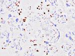
 in IHC (P)")
产品信息
Z2173MS
产品规格
种属反应
Human
宿主/亚型
Mouse
/ IgG2b, kappa
分类
Monoclonal
类型
Antibody
克隆号
KP10
抗原
Recombinant human p57Kip2 protein
偶联物
形式
Liquid
浓度
200 µg/mL
纯化类型
Protein A
保存液
tris with BSA, NP-40
内含物
<0.1% sodium azide
保存条件
4°C
运输条件
Ambient (domestic); Wet ice (international)
产品详细信息
A recommended positive control tissue for this product is Mole, however positive controls are not limited to this tissue type.
The primary antibody is intended for laboratory professional use in the detection of the corresponding protein in formalin-fixed, paraffin-embedded tissue stained in manual qualitative immunohistochemistry (IHC) testing. This antibody is intended to be used after the primary diagnosis of tumor has been made by conventional histopathology using non-immunological histochemical stains.
p57Kip2 (or CDKN1C) is a potent tight-binding inhibitor of several G1 cyclin complexes, and is a negative regulator of cell proliferation. The gene encoding human p57Kip2 is located on chromosome 11p15.5, a region implicated in both sporadic cancers, Wilm's tumor, and Beckwith-Wiedemann syndrome (BWS), a cancer syndrome, making it a tumor suppressor candidate. BWS is characterized by numerous growth abnormalities and an increased risk of childhood tumors. Several types of childhood tumors including Wilms' tumor, adrenocortical carcinoma and rhabdomyosarcoma display a specific loss of maternal 11p15 alleles, suggesting that genomic imprinting plays an important part. This region also contains two other imprinted genes, insulin-like growth factor II (IGF-II) and H19, both of which seem to be implicated in adrenal neoplasms.
Antibody is used with formalin-fixed and paraffin-embedded sections. Pretreatment of deparaffinized tissue with heat-induced epitope retrieval or enzymatic retrieval is recommended. In general, immunohistochemical (IHC) staining techniques allow for the visualization of antigens via the sequential application of a specific antibody to the antigen (primary antibody), a secondary antibody to the primary antibody (link antibody), an enzyme complex and a chromogenic substrate with interposed washing steps. The enzymatic activation of the chromogen results in a visible reaction product at the antigen site. Results are interpreted using a light microscope and aid in the differential diagnosis of pathophysiological processes, which may or may not be associated with a particular antigen.
A positive tissue control must be run with every staining procedure performed. This tissue may contain both positive and negative staining cells or tissue components and serve as both the positive and negative control tissue. External Positive control materials should be fresh autopsy/biopsy/surgical specimens fixed, processed and embedded as soon as possible in the same manner as the patient sample (s). Positive tissue controls are indicative of correctly prepared tissues and proper staining methods. The tissues used for the external positive control materials should be selected from the patient specimens with well-characterized low levels of the positive target activity that gives weak positive staining. The low level of positivity for external positive controls is designed to ensure detection of subtle changes in the primary antibody sensitivity from instability or problems with the staining methodology. A tissue with weak positive staining is more suitable for optimal quality control and for detecting minor levels of reagent degradation.
Internal or external negative control tissue may be used depending on the guidelines and policies that govern the organization to which the end user belongs to. The variety of cell types present in many tissue sections offers internal negative control sites, but this should be verified by the user. The components that do not stain should demonstrate the absence of specific staining, and provide an indication of non-specific background staining. If specific staining occurs in the negative tissue control sites, results with the patient specimens must be considered invalid.
靶标信息
This gene is part of a gene cluster on chromosome Xp11.23. The encoded protein contains a zinc finger motif often found in transcriptional regulators, however, its exact function is not known. Alternative splicing results in multiple transcript variants encoding the same protein.
仅用于科研。不用于诊断过程。未经明确授权不得转售。
篇参考文献 (0)
您是否在文献中引用过该产品?请点击下方按钮邮件告知我们。
生物信息学
蛋白别名: Beckwith Wiedemann syndrome (WBS); Cyclin dependent kinase inhibitor 1C (CDKN1C); Cyclin-dependent kinase inhibitor 1C; cyclin-dependent kinase inhibitor 1C (p57, Kip2); Cyclin-dependent kinase inhibitor p57; IMAGE; p57 Kip2; p57Kip2
基因别名: BWCR; BWS; CDKN1C; KIP2; p57; p57Kip2; WBS
UniProt ID: (Human) P49918
Entrez Gene ID: (Human) 1028
negative regulation of transcription from RNA polymerase II promoter
skeletal system development
kidney development
cell cycle arrest
regulation of mitotic cell cycle
aging
myeloid cell differentiation
adrenal gland development
positive regulation of transforming growth factor beta receptor signaling pathway
negative regulation of kinase activity
multicellular organism growth
negative regulation of phosphorylation
neuron maturation
camera-type eye development
negative regulation of transcription, DNA-templated
positive regulation of transcription, DNA-templated
negative regulation of epithelial cell proliferation
digestive system development
uterus development
embryonic placenta morphogenesis
genetic imprinting
negative regulation of protein serine/threonine kinase activity

