Search Thermo Fisher Scientific
EVOS S1000空间生物学成像分析系统简化了蛋白质空间定位的组织成像流程。
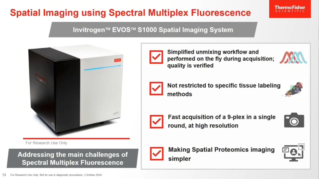
Invitrogen EVOS S1000空间生物学成像分析系统是一款宽场光谱成像系统,整合了多重光谱荧光、透射明场、相差及彩色明场成像等诸多功能。

核心特点:
- 快速:20分钟即可完成1 cm²组织的9标成像(10x;扫描、光谱解析及拼接)
- 灵活:兼容多种荧光染料、抗体、RNA探针及显色试剂
- 全面:支持光谱荧光、透射明场、相差及彩色明场成像
- 高灵敏度:配备420万像素16-bit科研级sCMOS相机,可清晰检测组织内复杂的多重染色信号
- 可变倍率:支持2.5x–40x物镜(5孔物镜转盘)
- 高效:自动化激光聚焦与软件辅助聚焦流程加速图像采集
- 易用:直观的软件界面,适用于不同经验水平的用户(图像采集、导航及数据检索)
- 兼容性:支持TIFF、OME-TIFF及分层图像格式,适配各类数据分析软件
- 高分辨率:光谱解析技术减少荧光串色,生成高分辨率图像

图1:使用EVOS S1000采集的扁桃体组织染色图像(透射明场图与彩色图)。
全面加速空间蛋白质组学研究
观看并了解多重成像技术在空间蛋白质研究中的最新进展。探索EVOS S1000如何简化工作流程、支持多染料应用并实现快速高分辨率成像。了解Invitrogen Aluora空间信号增强试剂如何通过高灵敏度和明亮信号提升抗体灵活性,以及如何利用荧光标记一抗实现快速多重染色。
用户评价
突破传统免疫组化限制
EVOS S1000空间成像系统助力从单一样本中获取更丰富的组织微环境信息。
- Why spatial proteomics?
- Spectral unmixing technology
- Benefits of multiplex IF
- Experimental workflow


Simplified tissue imaging workflow
Unlike existing technologies that can take several days to weeks, the EVOS S1000 instrument can complete the imaging process within several hours, with the greatest time savings found in its capability to generate a fully stitched, unmixed, multiplexed image for multiple samples. With the EVOS S1000 Spatial Imaging System, you have the flexibility to use a variety of labeling technologies, allowing you to select and utilize your preferred antibodies and reagents. The instrument outputs OME-TIFF files that are compatible with any third-party analysis software such as Halo and QuPath (QuPath: Open source software for digial pathology image analysis. Scientific Reports (2017).)

Figure 10: The spatial biology multiplex workflow incorporates the EVOS S1000 Spatial Imaging System that multiplexes more biomarkers with the use of spectral unmixing.
To build a panel for the EVOS S1000 Spatial Imaging system, start by choosing your targets, ensuring they align with your research goals. Next, create the panel by carefully picking out antibodies; use the Invitrogen SpectraViewer to determine where to place the 8 fluorophores labeling the antibodies plus DAPI. For the staining and labeling technology, use our Spatial Biology Reagent Selection Tool to chose what is right for your experiment. Finally, proceed with the imaging step to capture and analyze your samples.
Benefits of spectral unmixing technology


Antibodies and reagents


Ordering information
Resources and support


The Invitrogen EVOS S1000 Spatial Imaging System is only available in participating countries and regions including US, Canada, and Europe in 2024.
HALO is a trademark of Indica Labs, Inc.
For Research Use Only. Not for use in diagnostic procedures.